


Arthritis is a chronic disease that affects the connective tissue structure of the musculoskeletal system.The disease is characterized by a progressive process with gradual destruction of cartilage tissue.Arthritis is detected in most patients after the age of 65, since one of the causes of its development is the natural aging of the body.
The appearance of degenerative-dystrophic pathology is caused by previous injuries, endocrine and inflammatory diseases, excessive physical activity or, conversely, a sedentary lifestyle.The leading symptoms of arthritis are joint pain, swelling, and limited movement.
To diagnose pathology, instrumental studies are performed - radiography, arthroscopy, MRI, CT.Arthritis of severity 1 and 2 is treated conservatively with a course of medication, physical and massage therapy, and exercise therapy.In case of irreversible destructive changes in the joints, surgical intervention is indicated - arthroplasty, arthroscopy.
Pathogenesis mechanism
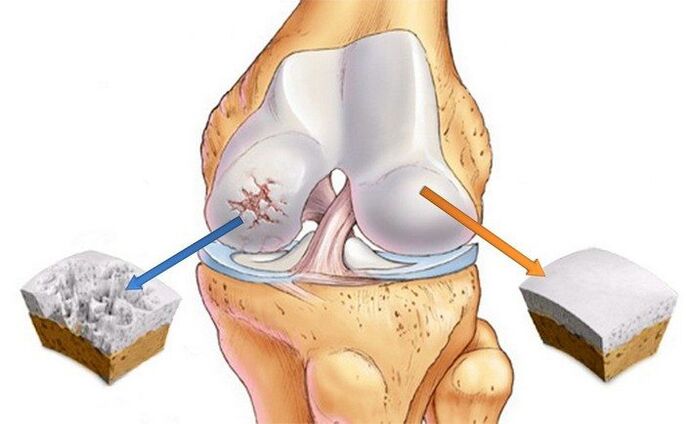
With arthritis, marked changes occur in the internal connective tissue structure.Deformative erosions form on cartilage tissues, causing destruction of collagen fibers, as well as proteoglycans including proteins (5–10%) and glycosaminoglycans (90–95%).As a result, the collagen network loses its stability and metalloproteinases begin to be released, destroying all types of extracellular matrix proteins.Destruction is accelerated by increased biosynthesis of collagenase and stromelysin.Normally, the normal quantitative values of enzymes are controlled by cytokines - small peptide messenger molecules.But as arthritis progresses, the concentration of these proteins decreases, which stimulates the release of large numbers of enzymes that damage cartilage tissue.
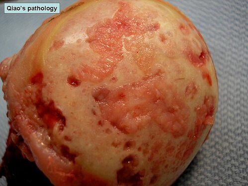
Proteoglycans with altered structures begin to absorb water molecules that they cannot retain.Therefore, excess fluid enters the collagen fibers.They “swell” and lose strength and elasticity.Negative changes also occur in the qualitative and quantitative composition of synovial fluid.When you have arthritis, the concentration of hyaluron in it will decrease.The hyaline cartilage no longer receives enough nutrients and oxygen to regenerate.Softening spots occur in the cartilage tissue, followed by cracks and specific necrotic developments.The bone ends are exposed and begin to undergo microscopic trauma when displaced relative to each other.
Causes and stimulating factors
The reasons for the development of primary (idiopathic) arthropathy are unknown.It occurs without any precipitating factors, so theories have been put forward about a genetic predisposition to premature cartilage destruction.Secondary arthritis develops as a result of other joint diseases or previous injuries.What can cause degenerative dystrophy:
- injury to the joint or nearby connective tissue structures - fracture, dislocation, damage to the meniscus, partial rupture of muscles, ligaments, tendons or complete separation from the bone base;
- congenital dysplastic disorders of joint development;
- disruption of endocrine glands, metabolic disorders;
- rheumatism or rheumatic fever;
- rheumatoid arthritis, reactive, metabolic, psoriasis or gout, polyarthritis;
- purulent arthritis caused by streptococci, epidermolysis bullosa or staphylococcus aureus;
- tuberculosis of any locality, brucellosis, chlamydia, gonorrhea, syphilis;
- degenerative diseases, for example, osteochondritis.
Hypermobility of the joints, due to special collagen production, predisposes to the development of joint disease.This condition is detected in 10% of the planet's inhabitants and is not considered pathological.But hypermobility is accompanied by weakness of the tendon-ligament apparatus, leading to frequent injuries, especially of the ankle joint (sprains, torn ligaments, dislocations).
Osteoarthritis is sometimes caused by blood disorders, such as hemophilia.Hemarthrosis, or hemorrhage into the joint cavity, causes deterioration of cartilage nutrition and its destruction.
Risk factors include old age, frequent loads on the joints beyond their strength limits, overweight, surgical intervention and hypothermia.

Risk groups include women in menopause, people living in unfavorable environmental conditions or exposed to toxic chemical compounds.If the diet lacks foods with vitamins and trace elements, it will create prerequisites for the gradual destruction of hyaline cartilage.
Clinical images
The danger of arthritis lies in the absence of symptoms at the first stages of development.The pathology manifests itself clinically gradually, the first signs appear against the background of significant destruction of cartilage tissue.Initially, a person feels mild pain with no clear localization.It appears after physical activity - lifting weights, doing sports.Sometimes the first clinical manifestation is a crunching or clicking sound when flexing or extending the joint.A person begins to notice that some movements are very difficult.However, in the early stages of arthritis, joint stiffness occurs in the morning and quickly disappears.
As the disease progresses, the patient also feels pain at night, not only causing sleep disturbances but also chronic fatigue.The severity of pain syndrome in the second stage increases with weather changes, exacerbation of chronic pathologies and acute respiratory viral infections.Range of motion is significantly reduced.The cause of joint stiffness is the thinning of cartilage, as well as the patient's conscious restriction of movement to avoid pain.This leads to increased load on the opposite joint, causing further damage.Arthritis is also characterized by other specific symptoms:
- pain causes spasms of skeletal muscles and the development of muscle contractures (limitation of passive movements in joints);
- crunching sounds in the joints, clicking sounds, clicking sounds when moving become continuous, occurring with almost any displacement of the bones relative to each other;
- muscle cramps often occur;
- Deformed joints lead to disorders of posture and gait;
- in the third stage of arthritis, the deformation is so pronounced that the joints are bent and the range of movement in them is significantly reduced or is completely absent;
- With grade 3 osteoarthritis of the knees, ankles, and hips, the patient must use a cane or crutches when moving.
In the absence of treatment, the pathology progresses, and during treatment, remissions are replaced by relapses and the frequency of exacerbations increases.Joint stiffness when exercising in the morning does not disappear for a long time but becomes permanent..
When examining a patient with grade 1 arthritis, the doctor noted only mild joint swelling and complete range of motion.In grade 2 disease, palpation shows pain and mild deformity.In the area of the joint space, bone thickening is observed.
Arthritis is characterized by the development of synovitis - an inflammatory process in the synovial membrane of the hip, knee, ankle and shoulder joints.Their main symptom is the formation of a circular seal in the joint area, when pressure is applied, fluid movement (oscillation) is felt.Acute synovitis may be accompanied by a temperature rise to 37-38°C, headaches and gastrointestinal disorders.
Diagnose
Diagnosis is made based on the results of instrumental studies, features of the clinical picture, medical history and patient complaints.General blood and urine tests do not give much information - all values remain within normal limits if the joint disease is not caused by a metabolic pathology.With the development of synovitis, the rate of erythrocyte sedimentation increases (30 mm/h), the level of leukocytes and fibrinogen in the blood increases.This indicates an acute or chronic inflammatory process occurring in the body.Changes in biochemical and immunological parameters occur in secondary forms of arthritis.
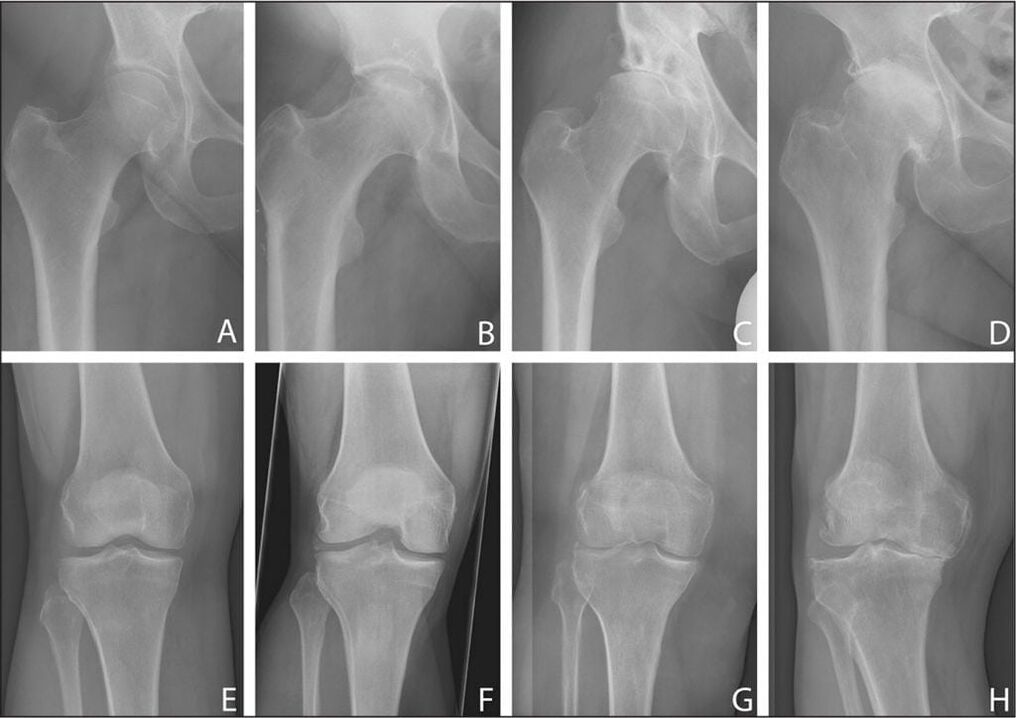
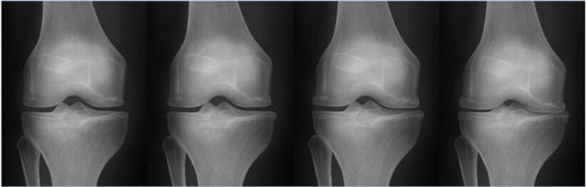
The most useful method for diagnosing degenerative-dystrophic pathology is radiography in frontal and lateral projections.
| Stages of osteoarthritis according to the Kellgren-Lawrence classification (1957) | X-ray signs of pathology |
|---|---|
| initial | There are no signs of radiation |
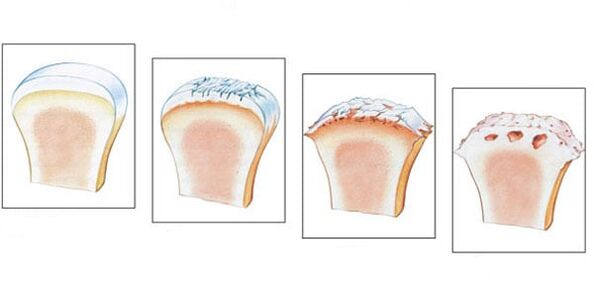
| Firstly | The joint space narrowing is not obvious and uneven.Slight flattening of the edges of the bone plates, formation of initial bone spurs or their absence |
| Monday | The joint space is noticeably narrowed, 2-3 times greater than normal, forming a large number of bone spurs and subchondral bone sclerosis.The appearance of cysts at the ends of bones |
| Tuesday | The appearance of marked subchondral osteosclerosis and large marginal osteophytes, significantly narrowing the joint space |
| Wednesday | Formation of large coarse bone spurs, almost complete fusion of the joint space, deformation and compaction of the ends of the bones that form the joint |
If after studying X-ray images, the doctor has doubts about the diagnosis, he will order a CT scan.And to evaluate the condition of connective tissue structures located near the joints, an MRI scan is performed.When using contrast agents, it is possible to dynamically assess the blood supply to tissues and determine the stage of the inflammatory process in the development of synovitis.
Basic treatment methods
Arthritis is still an incurable disease because there is no pharmacological drug to regenerate cartilage tissue.The main goal of therapy is to stop the progression of the pathology and maintain joint mobility.Long-term, complex treatment, using both topical and systemic medications.The patient should avoid severe stress on the joints and, if necessary, limit the range of motion with orthopedic devices - orthotics, elastic bandages.Overweight patients need to adjust their diet to gradually reduce body weight and follow a diet.
After the disease is in stable remission, the patient receives daily physical therapy.The first training sessions are performed under the guidance of a physical therapist, after which the patient performs a series of exercises at home.Exercise therapy can be supplemented with swimming, yoga and cycling.
To reduce the severity of pain, drugs of various clinical and pharmacological groups are prescribed:
- Non-steroidal anti-inflammatory drugs in the form of ointments, tablets, intravenous solutions with active ingredients;
- Intra-articular injection of anesthetic solutions combined with glucocorticosteroids;
- muscle relaxants to eliminate muscle spasms and restrictive spasms.
The treatment regimen includes B vitamins, sedatives and, if necessary, sedatives and antidepressants.Chondroprotectors are essential for long-term use.This is the only group of drugs capable of partially restoring cartilage tissue.
To enhance their clinical activity, physiotherapeutic procedures are performed - laser therapy, magnetic field, UHF therapy.
Any pain in the joints is a signal to immediately consult a doctor.Therapy performed in the early stages of joint disease will prevent cartilage destruction and avoid loss of function and disability.